Besides being in the office 8-5pm, I spent Monday and Tuesday evening studying cell biology, last night I attended a lameness workshop at Nantwich Equine Vets and this evening I have ridden my friends pony Toby who has a bit of a separation issue when split up from his field mate Tigger. So to say this evenings ride was eventful would be an understatement!
This week's post is going to be based around the workshop I attended last night:


 |
| Can't beat an information pack! |
The Equine Lameness Examination (What the vet will do and why)
"A lameness examination is a forensic investigation of the horse."
- Alasdair Topp BVM&S Cert AVP (EOS) MRCVS
History
First of all the vet will want to know about the horses history. Key things your vet will ask are:
- Age
- Sex
- Breed
- Use/Performance level
- Management - Surfaces worked on/stabled or at grass etc.
- Previous problems?
- Duration of lameness
- What does the owner think is wrong
Observation
Next your vet will want to spend a few minutes just observing your horse all over. Things a vet will look at include:
1.Confirmation
- How are his forelimbs? Are they in proportion to the rest of his body? Are his feet turned out or pigeon toed? His knees - are they over at the knee/Back of the knee? - Horses that are back at the knee can have excessive amounts of strain placed on the tendons
- How are his hind limbs? Are they well put together? Is he base narrow or base wide? His hocks? Are they sickle/over straight/up in the air?
- His back - is he roach backed or have a dipped back?
- His body - the shoulder and rump angle should be 90° anything over this and the horses strides will tend to be shorter and anything under will result in a weaker structure.
2. Foot balance
3. Symmetry of the horse
4. Swelling - bony/soft tissue
5. Posture - e.g. laminitic ponies will tend to rest on the heels of all four feet
3. Symmetry of the horse
4. Swelling - bony/soft tissue
5. Posture - e.g. laminitic ponies will tend to rest on the heels of all four feet
6. Distribution of weight placed over limbs
 |
| Image - A Photographic Guide to Confirmation - Such a good read! |
Palpation
This is where your vet will examine the horse for heat, swelling and pain.
Usually starting at the foot using hoof testers and working the way up the limb using his hand, your vet will feel for any heat/swelling and locate areas of causing pain. He may do this by applying a slight pressure in certain areas to see the reaction of the horse.
After viewing the limbs your vet may want to view the range of movement in the spine.
- Ventroflexion is the arching of the spine, to do this the vet will either use his hand or run a pen ventrally along the abdomen so the horse arches his spine.
- Dorsiflexion is the hollowing away from pressure applied either side of the withers.
- Lateral flexion is a sideways flexion. By running a hand down the spine applying pressure to either side of the spine should cause the horse to curve away in lateral flexion.
The overall body will also be looked at to see if there is any muscle wastage/atrophy or unusual sweat marks.
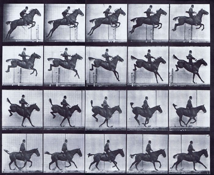
Movement
Your vet will use a baseline for each lameness examination. This is basically a starting point and when viewing the movement of the horse, each issue will have its own baseline.
Your vet will want to:
- View the horse walking and trotting in a straight line. Things your vet will look for when doing this are head nods and hip action.
- Flexion tests - using reasonable force for about 40 seconds the joint in question is flexed to full capacity to accentuate any problems.
- Extension tests - joints may be extended as opposed to flexed, this is rarely used.
- Lunge on a hard surface - this can highlight bone related issues and typically shows inside limb lameness.
- Lunge on a soft surface - this can highlight soft tissue issues and typically shows outside limb lamness.
- Ridden - some horses may only show lameness when ridden so your vet will want to see this.
 |
| Lunging Crunchie |
Things your vet may notice (and you too!):
- Forelimb lameness - a head nod on the sound leg
- Hind limb lameness - a hip hike on the lame limb and a possible head nod on the contralateral forelimb (opposite forelimb) when this occurs it's easy to mistake hind limb lameness for forelimb lameness
- Hoof pattern sound - unusual pattern caused by a toe drag?
- Change in limb flight during cranial and caudal phases - as the limb moves forward (cranial phase) it may have a shorter stride due to lameness
- Foot landings
- Mechanical lameness - physically abnormality preventing the a normal motion
Your vet will then grade the lameness on a scale of 0-5.
0 - No lameness found
1 - Mild
2 - Obvious
3 - Pronounced
4 - Severe
5 - Not weight baring/inability to move
Diagnostic Modalities
This is where the vet will now try to localise the lameness to a specific region of the body. This can be done by using one of the below modalities:
Nerve Blocks – The most common modality used in localising lameness. Post trot up/flexion test, a vet would have identified a limb in which he believes the lameness is occurring from and starting from the foot and working his way up the limb, he would inject local anaesthetic to block the nerves until the lameness was eliminated.
Magnetic Resonance Imaging (MRI) – Using magnetic fields and radio waves, this technology can provide a clear image of what’s going on under the surface of the skin. It's good for areas like cartilage.
Nuclear Scintigraphy – This requires the horse to be kept in for a few nights at a veterinarian practice due to the use of radioactive chemicals which are injected into the body and are then detected from outside to see where the most chemical build up occurs. This would highlight any areas that may need investigation.
Arthroscopy – A diagnostic tool by where a surgical procedure is required in the form of keyhole surgery to look into the joints of a horse. This is done using a very thin telescope and a light source.
Radiography (x-ray) – Provides images on the skeletal structure by using electromagnetic radiation. The horse may need an additional scan should the x-ray prove to be inconclusive in its findings.
Ultrasound scan – Produces images of the fibre patterns in the tendons, ligaments, tissues, vessels, organs and fluid accumulation by using sound waves. Veterinarian practices also use ultrasound as a guidance tool when medicating certain areas of the body.
Thermography - Using an infrared camera, the technician has the ability to capture real time heat maps in the form of a thermogram. Thermograms are pictorial representations of the surface temperature of the horse in question. Measuring the surface temperature of the horse gives us an indication of any changes in the vascular, muscular, skeletal and nervous systems as we are able to view any inflammation going on underneath the skin which is undetectable to the human hand or eye.
Hopefully by using one of the above modalities you will have been able to locate the problem and the right course of treatment will be then arranged by your vet.
I will be attending another workshop next month on common forelimb lameness and so I will keep you posted on how that goes too!
Jess x

.jpg)